Tán sỏi laser sỏi trong túi thừa niệu đạo ở nữ giới
Túi thừa niệu đạo rất hiếm gặp ở nữ giới gây các biến chứng nhiễm trùng tiết niệu, sỏi trong túi thừa. Sử dụng Laser tán sỏi có những ưu điểm: ít đau, thẩm mỹ, nhanh ra viên,
| Int Urogynecol J (1995) 6:34-36 9 1995 The International Urogynecology Journal | International Urogynecology Journal |
Case Report
Transurethral Laser Lithotripsy in a Female Urethral Diverticulum Containing Stones
M. N0hr and S. Walter Department of Urology, Aalborg Sygehus, Aalborg, Denmark
| Abstract: A case of transurethral laser lithotripsy of a large stone in a female urethral diverticulum is presented. It is concluded that the technique is more preservative to the urethra and sphincter than conventional surgery. Keywords: Endoscopic technique; Laser lithotripsy; Stress urinary incontinence; Urethral diverticulum; Urolithiasis Introduction Transurethral techniques applied to the surgical treatment of urethral diverticula in females have been reviewed by Miskowiak [1]. We present a case of transurethral laser-induced lithotripsy in urethral diverticula complicated by Case Report A 41-year-old woman was referred to investigation of recurrent lower urinary tract infections. In addition, she claimed periodic stress urinary incontinence and interruption of the stream during urination. Intravenous urography combined with a urethrogram showed diverticula with stony shadows on both sides of the urethra; Correspondence and offprint requests to: Dr Mads N0hr, Lindenborgvej 53, DK-9200 Aalborg SV, Denmark. | each of the calculi measured i cm in diameter (Fig. 1). A physical examination, including urethral palpation, revealed a hard mass on both sides of the urethra. Transurethral endoscopy showed a narrow diverticular ostium on each side of the midurethra. Both diverticula contained a large stone, but there were no signs of pus or neoplastic growths. The proximal and distal urethra, the bladder neck and the bladder were normal. Even after a careful incisional opening of the diverticular ostium in the lateral direction with Sachse's urethrotome, it was not possible to remove the stones because of their size. To avoid damage to the sphincter, further incision was not performed. During a 13-day period the patient received laser-induced lithotripsy under general anesthesia five times. Under optical control, pulsed d~.e laser impulses were fired at the stones (Pulsolith ~, Technomed International, Paris France) [2,3]. The stones were successfully disintegrated and the residual small fragments were removed with biopsy forceps or passed spontaneously. A plain X-ray of the region showed no residual calculi. Clinically the patient recovered fully. She had no symptoms of urinary tract infection or stress urinary incontinence. Pre- and postoperative urodynamic studies showed improved maximal flow rate and average flow rate, with equal amounts of voided volumes after the treatments. Stone analysis showed a calcium-magnesium-ammonium-phosphate composition most likely caused by infection. Over the next 24 months the patient was followed with urethrocystoscopy and urinary bacterial analysis. In one of the diverticula there was recurrence of a few stones and a mild urinary tract infection. The stones were removed and since then the patient has been asymptomatic, without stones, infections or neoplastic growths in the diverticula. |
Transurethral Laser Lithotripsy in a Female Urethral Diverticulum Containing Stones
Discussion: Female urethral diverticula may cause repeated urinary tract infections and stress urinary incontinence as in this case. In addition, the patient may complain of dysuria, urgency, frequency, dribbling after voiding, dyspareunia and urethral discharge. Diverticula containing stones may cause further discomfort, including urinary retention, interrupted urinary stream and urethral bleeding. Physical examination with urethral palpation almost always gives the diagnosis, but should be verified with a plain X-ray, a urethrogram and direct visualization by urethroscopy. Transvaginal diverticulectomy may be performed. When inflammation is present the operation should be in two stages, with a primary incision and a secondary excision. Surgery of the urethra may cause bladder | injury, fistula formation, lesions of the bladder neck, sphincter damage and urethral strictures. Furthermore, the recurrence rate of the diverticula following excision is up to 18%-20% [4,5]. The transurethral treatment of urethral diverticula is directed mainly towards drainage by internal incision of the ostium [1], but when complicated by large calculi this treatment may be insufficient. In our case the calculi were too large and required wide incisions, with risk of damage to the bladder neck and the sphincter. Urethral pressure profiles to locate the high-pressure zone may be useful when wide incisions are required. Pressure profiles were not performed in our case, and we managed the problem by performing a small internal incision preserving the sphincter and, following the laser-induced disintegration of the calculi, they were removed with biopsy forceps or passed spontaneously. The drainage of the diverticulum may result in a reduction in size due to decreased intraluminal pressure, and thereby give relief of symptoms. Lithotripsy with a thin ultrasound probe through the endoscope may also be useful [6,7]. Shock-wave lithotripsy with ultrasound may be difficult or even impossible in this region, and we have no experience with it. We chose to try laser lithotripsy because we have had a good experience with it in other regions. The pulsed dye laser technique and fragmentation of ureteral calculi is described in other papers [3,8,9]. In contrast to conventional surgical techniques, the transurethral techniques for managing calculi may require a large number of repeat treatments under general/regional anesthesia. The laser probe used in our department at that time was constructed for use in a small-caliber ureteroscope and not for the urethroscope, which had a large working channel. As a result, it was difficult to get the probe in contact with the calculi. Since then we have acquired better equipment, but this explains the need for repeat treatments [9]. As lon~ as repeated treatment shows progress, the patient may accept the procedure. On the other hand, the transurethral technique is more preservative to the urethra and sphincter and shortens the hospital stay [1]. These patients should always be followed up because of the increased risk of developing adenocarcinoma in the remaining diverticula [4]. Any sign of developing adenocarcinoma should lead to excision of the diverticulum. If the transurethral procedure is not accepted by the patient, marsupialization may be suggested instead. References 1. Miskowiak J. Surgical treatment ofurethral diverticula in females, with focus on transurethral techniques. Int Urogynecol J 1991;2:112-114 2. Beck EM, Vaughan ED Jr., Sosa RE. The pulsed dye laser in the treatment of ureteral calculi. Semin Urol 1989;7:25-29 3. Juhl Jensen V, Krarup T, Walter S. Pulsed dye laser treatment of ureteral calculi. Scand J Urol Nephrol Suppl 1991;138:31-33 36 M |
| 4. Ginsburg DS, Genadry R. Suburethral diverticulum in the female. Obstet Gynecol Surv 1984;39:1-7 5. Lichtman AS, Robertson JR. Suburethral diverticula treated by marsupialization. Obstet Gynecol 1976;47:203-206 6. Durazi MH, Samiei MR. Ultrasonic fragmentation in the treatment of male urethral calculi. Br J Urol 1988;62:443-444 7. Sharfi AR. Presentation and management of urethral calculi. Br J Urol 1991 ;68:271-272 8. Dretler SP, Watson G, Parrish JA, Murray S. Pulsed dye laser fragmentation of ureteral calculi: initial clinical experience. J Urol 1987;137:386-389 9. Anson K, Seenivasagam K, Miller R, Watson G. The role of lasers in urology. Br J Urol 1994;73:225-230. 9. Anson K, Seenivasagam K, Miller R, Watson G. The role of lasers in urology. Br J Urol 1994;73:225-230 | EDITORIAL COMMENT: This is the first instance of treatment of a urethral diverticular stone with laser lithotripsy. The presence of a stone in a diverticulum is an uncommon occurrence and certainly difficult to remove without disruption of urethral integrity in one way or another. If preservation of the urethra at all costs is the goal of the therapy, then this is one treatment that can be added to our armentarium. With better equipment than used in this case, multiple treatments can be avoided. It is important to remember that carcinoma may occur in the diverticulum as well, and that any diverticulum left behind must be periodically assessed for this possibility |
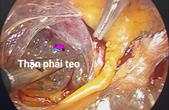
Hình ảnh tán sỏi trong túi thừa niệu đạo ở nữ giới tại Bv ĐHY Hà nội
Các bài viết liên quan:
1. Tán sỏi niệu đạo - Sỏi niệu đạo nam giới
2. Phẫu thuật tán sỏi niệu quản nội soi ngược dòng bằng Laser
3. Các nguyên nhân điều trị sỏi niệu quản nội khoa thất bại
4. Sỏi niệu quản nhỏ, kẻ giết người thầm lặn
5. Sỏi niệu đạo nam giới
Tin nổi bật
- GIẢI PHẪU ĐƯỜNG TIẾT NIỆU TRÊN ỨNG DỤNG TRONG NỘI SOI NIỆU QUẢN - THẬN NGƯỢC DÒNG
10/08/2023 - 21:22:35
- Hướng dẫn các bước phẫu thuật điều trị tràn dịch màng tinh hoàn ở người trưởng thành
16/07/2023 - 22:11:23
- Các bước phẫu thuật nội soi sau phúc mạc cắt thận mất chức năng
08/07/2023 - 18:24:37
- Các bước phẫu thuật nội soi sau phúc mạc cắt thận mất chức năng
08/07/2023 - 18:07:24
- MỔ MỞ ĐIỀU TRỊ THOÁT VỊ BẸN - THOÁT VỊ BẸN NGHẸT Ở TRẺ EM
20/12/2021 - 16:23:17
- Một số phẫu thuật điều trị bệnh lý ở tinh hoàn
12/12/2021 - 15:52:47


.jpg)


